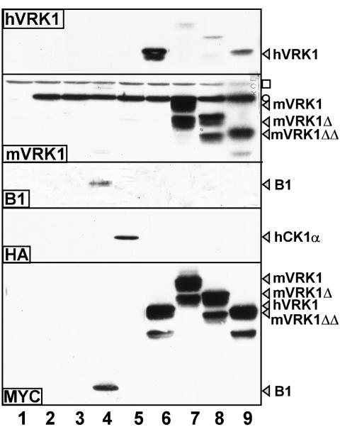
FIG. 3.
hVRK1, mVRK1s, hCK1α, and wt B1 are expressed during infections performed with the ts 2 recombinant viruses. BSC40 cells were either left uninfected (lane 1) or infected with wt virus (lane 2), ts2 (lane 3), ts2/B1 (lane 4), ts2/hCK1α (lane 5), ts2/hVRK1 (lane 6), ts2/mVRK1 (lane 7), ts2/mVRK1Δ (lane 8), or ts2/mVRK1ΔΔ (lane 9) at 31.5°C for 17 h (MOI of 10). Cell lysates were subjected to immunoblot analysis with antisera directed against hVRK1, mVRK1, B1, HA, or c-MYC followed by incubation with an HRP-conjugated secondary antibody and chemiluminescent development. In the hVRK1 panel, a strong signal was seen in cells infected with the ts2/hVRK1 recombinant; in addition, the sera show a moderate level of cross-reactivity with the mVRK1 proteins but do not detect endogenous hVRK1 levels. Two cross-reactive proteins (one viral, ○, and one cellular, □) are seen in the mVRK1 panel. For the blot developed with anti-B1 serum, endogenous levels of B1 are not detected, but the B1 protein overexpressed in the ts2/B1 recombinant is easily seen.

