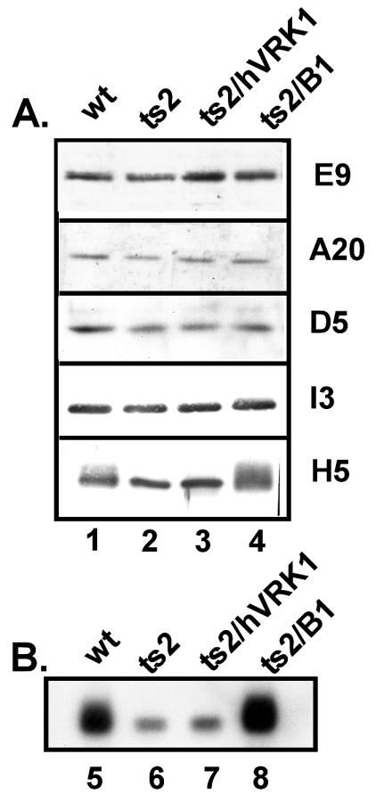
FIG. 5.
Does the ts2 defect or its rescue affect the accumulation or phosphorylation of viral replication factors? (A) Levels of replication proteins are not affected by the presence or absence of active B1. Confluent monolayers of BSC40 cells were infected with wt virus (lane 1), ts2 (lane 2), ts2/hVRK1 (lane 3), or ts2/B1 (lane 4) (MOI of 15) and incubated at 39.7°C for 4 h prior to harvesting. Cell lysates were fractionated on SDS-12% polyacrylamide gels, and proteins were transferred to nitrocellulose filters, which were cut appropriately and probed with antibodies directed against E9, A20, D5, H5, or I3. After incubation with HRP-conjugated secondary antisera, immunoreactive proteins were visualized either by colorimetric (H5 and I3) or chemiluminescent (E9, A20, and D5) development. Equivalent levels of all five proteins are seen in the four samples; note, however, that the H5 protein seen in lanes 1 and 4 is heterogeneous, containing additional species with reduced mobility. (B) Immunoprecipitation of 32P-labeled H5 protein: H5 hyperphosphorylation is B1 dependent and is not required for the rescue of DNA replication. Confluent 35-mm-diameter dishes of BSC40 cells were infected with wt virus (lane 5), ts2 (lane 6), ts2/hVRK1 (lane 7), or ts2/B1 (lane 8) (MOI of 15) and incubated at 39.7°C for 3 h. Cells were then incubated in phosphate-free medium supplemented with 100 μCi of 32PPi per ml at 39.7°C for 2 h prior to harvesting. Radiolabeled cell lysates were immunoprecipitated with anti-H5 serum, and immunocomplexes were fractionated on an SDS-12% polyacrylamide gel and visualized by autoradiography.

