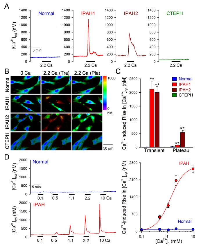
Figure 1. Enhancement of extracellular Ca2+-induced increase in [Ca2+]cyt in PASMC from IPAH patients.
A-C. Representative records (A), pseudo-color images (B), and summarized data (means±SE, C) showing extracellular Ca2+-mediated changes in [Ca2+]cyt in PASMC from normal subjects (n=45 cells), IPAH patients (n=160 cells), and CTEPH patients (n=27 cells). Two kinetically different responses of [Ca2+]cyt to extracellular Ca2+ in PASMC from two IPAH patients are shown in the middle panels. D. Representative traces of [Ca2+]cyt changes in response to extracellular application of 0.1, 0.5, 1.1, 2.2, and 10 mM Ca2+ (left panels) and the dose-response curves (right panel) in normal PASMC (upper panel) and IPAH-PASMC (lower panel). The EC50 for extracellular Ca2+-induced [Ca2+]cyt increase in IPAH-PASMC is 1.22 mM.

