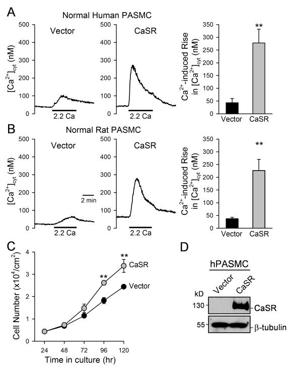
Figure 5. Overexpression of CaSR in normal PASMC enhances extracellular Ca2+-induced [Ca2+]cyt increase and promotes cell proliferation.
A and B. Representative records of [Ca2+]cyt changes (left panels) and summarized data (means±SE, right panels) extracellular Ca2+-induced [Ca2+]cyt increases in human (A) and rat (B) PASMC transfected with an empty vector (Vector, n=12) and the human CaSR cDNA (CaSR, n=8). C. Summarized data (means±SE) showing the total numbers of normal human PASMC transfected with the vector (solid circles) and CaSR gene (grey circles) after incubation in growth media for 24, 48, 72, 96, and 120 hrs. The growth curves for vector control PASMC and IPAH PASMC are significantly different (P<0.01, n=3 experiments). **P<0.01 vs. vector. D. Western blot analysis on CaSR in human PASMC transfected with an empty vector (Vector) or CaSR.

