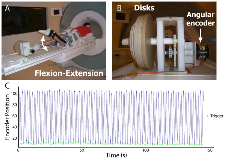
Figure 1. Experimental setup.
Subjects were positioned in the head-first, prone position in an MR-compatible exercise device during repeated knee flexion and extension (subject pictured outside the scanner) (A). Cyclic rotation of inertial disks resulted in active lengthening of the biceps femoris muscle (Silder et al., 2009), while an angular encoder signal was sent to a laptop computer running LabVIEW (B). Encoder position values (line) were used to trigger the scanner (asterisk) to begin image acquisition at the onset of knee extension (C). (Example data are from a single DENSE acquisition.)

