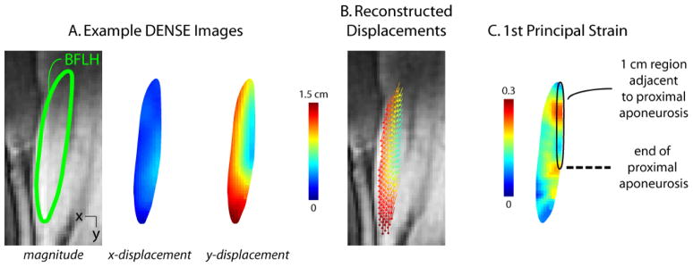
Figure 3. Example DENSE images, reconstructed displacements and strain map.

Displacement encoding with stimulated echoes (DENSE) images were acquired in an oblique-coronal plane containing the biceps femoris long head muscle (A). Measured displacements were used to reconstruct time-varying tissue position at a pixel-wise resolution (B), where vectors represent displacement from the first image and the vector’s color represents the magnitude of displacement. First principal strain was defined as the most positive eigenvalue of the Lagrangian strain tensor and was averaged in the region within approximately 1 cm of the proximal aponeurosis (C).
