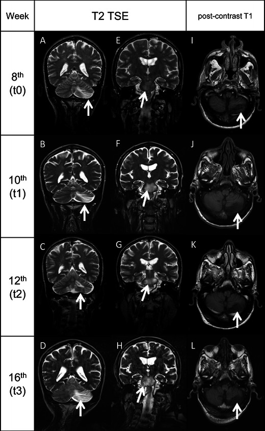
Fig. 2.

MRI evolution of the brain lesions. A–D, T2-TSE sequences showing the evolution of the left cerebellar lobe lesion (arrows) from t0 to t3. E–H, T2-TSE sequences showing the evolution of the lesion of the pons (arrows) from t0 to t3. No mass effect was seen in all the images collected. I–L, post-contrast T1-weighted sequences demonstrating the absence of contrast enhancement (arrows) in the left cerebellar lobe lesion (MRI). All the white matter lesions did not show enhancement in T1-weighted sequences after contrast administration. The lack of mass effect and contrast enhancement in all the MRI performed from t0 to t3 reduced the reliability of an IRIS-PML, which is usually characterised by a strong inflammatory infiltration of tissues, determining an alteration of blood–brain barrier which translates in penetration of contrast inside the lesions and oedema, which is the cause of mass effect
