Fig. 1.
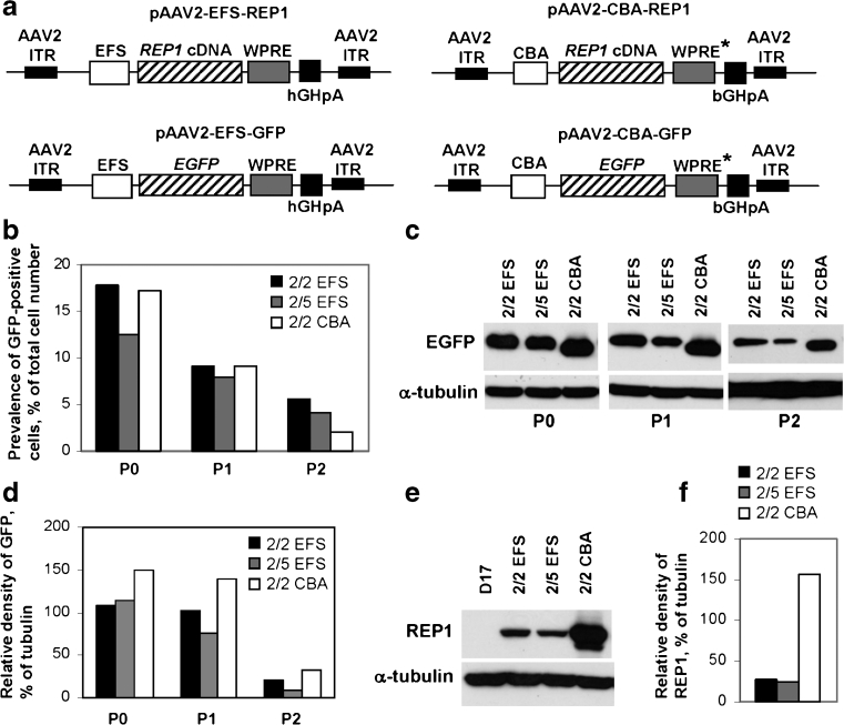
Expression of CHM/REP1 cDNA and EGFP transgenes in dog D17 cell line. a Plasmids pAAV2-EFS-REP1 and pAAV2-CBA-REP1 carry the CHM/REP1 cDNA transgene with Kozak sequence at the 5′-end. Plasmids pAAV2-EFS-GFP and pAAV2-CBA-GFP were used to generate control viral vectors. WPRE* is a WPRE that has been modified by deleting the We2 promoter/enhancer and mutating the We1 promoter (for other details, see “Materials and methods”). b–d D17 cells were transduced with AAV2/2-EFS-GFP, AAV2/5-EFS-GFP and AAV2/2-CBA-GFP. Expression of GFP was analysed 2 days post-transduction (P0). Cells were replated, cultured for additional 6 days and analysed (P1), then replated and analysed after 4 days in culture (P2). b FACS analysis shows prevalence of transduced cells (% of total cell number). c Immunoblot analysis of the total protein lysate using GFP antibody and α-tubulin antibody as a loading control. d Quantification of the western blot shown in c using ImageJ software. Data are presented as relative density of GFP signal in relation to α-tubulin signal. e Immunoblot analysis of the D17 cells transduced with AAV2/2-EFS-REP1, AAV2/5-EFS-REP1 and AAV2/2-CBA-REP1 using 2F1 antibody specific for human REP1 and α-tubulin antibody as a loading control. Cells were analysed 48 h post-transduction. f Quantification of the western blot shown in e using ImageJ software. Data are presented as relative density of GFP signal in relation to α-tubulin signal

