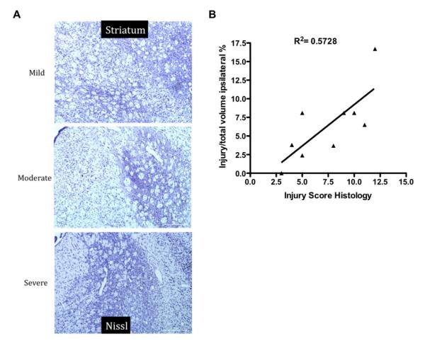
Figure 3.
T2-weighted imaging during subchronic injury predicts histological outcome 2 weeks after middle cerebral arterial occlusion at P10. (A) Representative Nissl-stained images of the striatum at P25. (B) A regression analysis comparing injury identified noninvasively on T2-weighted imaging to histological injury score revealed a significant correlation between these 2 parameters (D). (n = 9, R2 = 0.5728, P < .02).

