
Figure 1.
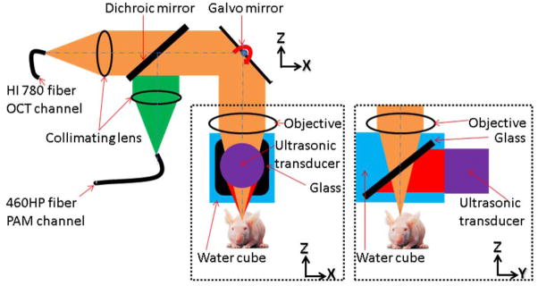
Schematic of the dual-modality OCT/PAM imaging probe: The OCT system was connected to the dual-modality probe through a single-mode fiber (HI-780, Thorlabs). The 532 nm pulsed laser was connected to the probe with another single-mode fiber (460-HP, Thorlabs). The collimated OCT sample and the 532 nm PAM excitation beams were combined using a dichroic mirror and focused by a microscope objective (NA 0.1) at 200 μm below the tissue surface. Fast optical scanning along one axis (B-scan) was done by a galvanometer mirror. Upon the absorption of the laser pulse by the tissue, photoacoustic waves were generated. Thereafter, the waves were reflected by a glass plate placed at 45 degrees between the objective and the animal, and detected by the cylindrically focused ultrasonic transducer (GE, 25 MHz bandwidth). The whole probe was attached to a one-dimensional mechanical stage, which scanned perpendicularly to the fast optical scan axis. The OCT A-line image and the PAM A-line image were acquired sequentially for each A-line.