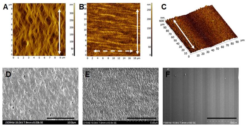
Fig. 1. Characterization of the nanofibrillar collagen membranes.
Characterization of the fibril diameter (A, 100 nm; B, 30 nm) and microgroove size (C, 60-μm wide microgrooves and 100 nm fibrils) by atomic force microscopy. Solid arrow shows the direction of the fibrils, and dashed arrow shows the direction of the crimp. Visualization of crimped fibrils (D, 100 nm and E, 30 nm) and microgrooves (F, 60-μm wide microgrooves and 100 nm fibrils) by scanning electron microscopy. Scale bar: 10 μm (D), 5 μm (E), 500 μm (F).

