
Figure 2.
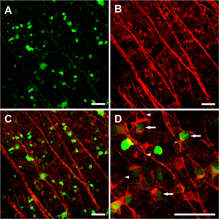
Fluorescent photomicrographs showing characteristics of green fluorescent protein (GFP)+ transfected cells and βIII tubulin+ surviving retinal ganglion cells (RGCs) in the same field of retinal whole mount. Five weeks after intravitreous injection of 5.0 μl viral vectors, extensive GFP expression mostly in the RGC layer was shown (A). B shows βIII tubulin+ surviving RGCs in the same field. Merged figure (C) of A and B showing many GFP+ cells are also βIII tubulin+ and are therefore retinal ganglion cells. D: Arrows show examples of transduced RGCs, arrow heads show non-transduced RGCs. Scale bar, 50 μm.