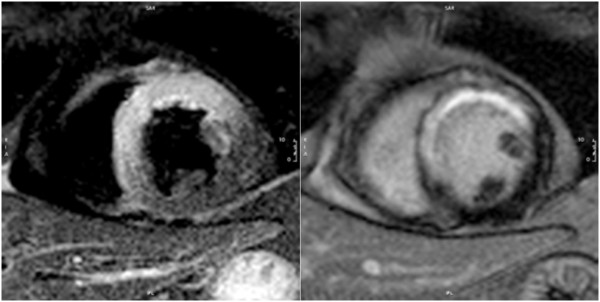
Figure 5.

CMR in acute myocardial infarction. Acute reperfused infarction of the left anterior descending artery territory. Left: T2-weighted image (short-tau inversion recovery, STIR) in a midventricular short axis view with increased SI in the affected segments. Right: LGE image in the same orientation.
