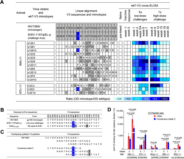
Figure 2.
Mapping of anti-V3 binding Abs. A. Sequences of recombinant phages were assigned to V3 crown of HIV1084i or SHIV-1157ipEL-p. Gray shading, linear homologies; blue shading/white letters, amino acid difference between the two HIV-C envelopes (M307I). V3 mimotopes isolated using week 7 plasma (wk7-V3 mimotopes) were tested for plasma Ab binding using 14 time points of RRi-11 and RTr-11. Negative control, pre-immune plasma; weeks −1 and 0, vaccine-induced Ab responses; weeks 1–7, also include Ab responses induced during low-dose virus challenges; weeks 8–30, Ab responses after all challenges. Data illustrate results from two independent assays. Binding patterns are shown in form of a heat-map. OD signals 3x higher than signals detected with the wildtype phage control were considered positive. B. V3 amino-acid sequences of HIV1084i, SHIV-1157ipEL-p and SHIVSF162P4. The two HIV clade C envelopes of SHIV-1157ipEL-p and HIV1084i differ in only one residue in the V3 crown (M307I; HXB2 numbering scheme [17]; highlighted in blue). SHIVSF162P4 shares the same gp120-sequence as HIVSF162[18] and has three mutations compared with the 1084i immunogen (M307I (blue), and R308P and Q315K (gray)). C. Sequences of synthetic peptide sets used for plasma Ab titration and peptide absorption analysis. Peptides corresponding to consensus clade C sequence differed in two amino-acid residues compared with immunogen-related peptides (HIV1084i): M307I (blue) and A316T (gray). D. Plasma samples of five vaccinees [12] were assessed for binding Ab specificities at weeks 0 and 7 using two different V3 peptide sets (red bars, 1084i; blue bars, consensus clade C). Height of each bar, average titer calculated from three independent assays; error bars, standard error of the mean (SEM). Statistically significant differences between weeks 0 and 7 are indicated (if P < 0.05).

