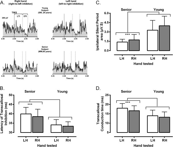
Figure 1.
Examples of ispsilateral inhibition and mean group differences. A. Examples of rectified and averaged EMG traces (n=5 trials) depicting ipsilateral silent periods elicited in response to transcranial magnetic stimulation (TMS) in two participants, young and senior. As illustrated, several measures of transcallosal inhibition could be derived from iSP recordings. The latency of transcallosal inhibition (LTI) was computed as the time interval between the cortical stimulus (thick dotted line) and the iSP onset (first thin dotted line), which was determined as the first sustained decline in EMG activity when compared to mean pre-stimulus level (horizontal dotted line). The duration of transcallosal inhibition (DTI) was estimated as the time interval between the iSP onset and offset (second thin dotted line), which as determined as the point where the EMG activity returned to pre-stimulus level. Finally, the depth of ispsilateral inhibition was estimated by calculating the iSP area, as depicted by the blank area delimited by the two vertical dotted lines (DTI) and below the horizontal line in the recordings. Note that timing measurements are given only for illustrative purposes, as the real estimates were derived from a trial-by-trial analysis. B. C and D. Mean variations (± 1 SD) in measures of ispsilateral inhibition (B, LTI: Latency of transcallosal inhibition; C, iSP area: ispsilateral silent period area, D, TCT: transcallosal conduction time) derived from each hand/ hemisphere for the two groups of participants. Note again the major differences between age groups as indicated by the asterisks (p<0.001).

