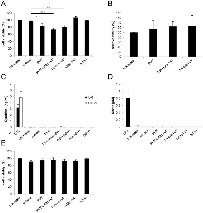
Figure 3. Effect of PrP106-126 A117V on microglia.
Following treatment for 24 hours with 10 µM PrP106-126 A117V with or without nAbs-PrP or ft-PrP, the MTT assay was performed to measure the mitochondrial activity of microglial cells. Values are normalized to untreated cells (A). The vitality of treated cells was verified by staining microglia with fluorescein diacetate/propidium iodide, and values are normalized to untreated cells (B). Supernatants of the cells were subjected to cytokine ELISA (C) and Griess assay (D) with LPS (1 µg/ml) as the positive control. (E) Conditioned supernatant of microglial cells was administered to primary neurons for 24 hours, and the MTT assay was performed. Values are normalized to untreated cells. All experiments were performed at least three times independently.

