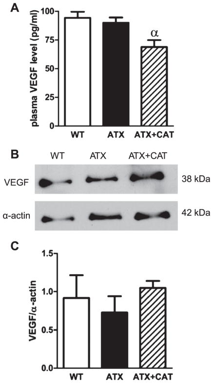
Fig. 6.
A: effect of a chronic treatment of 6-mo LDLr−/−:hApoB+/+ (ATX) mice with CAT on plasma VEGF level. B: representative example of Western blot illustrating VEGF protein expression of total cerebral vessels from WT and ATX mice treated, or not, with CAT. C: graph represents the protein expression normalized to a reference sample (loaded in every experiment) and after normalization with smooth muscle α-actin. Values are means ± SE of 4 –5 mice. αP < 0.05 vs. ATX mice.

