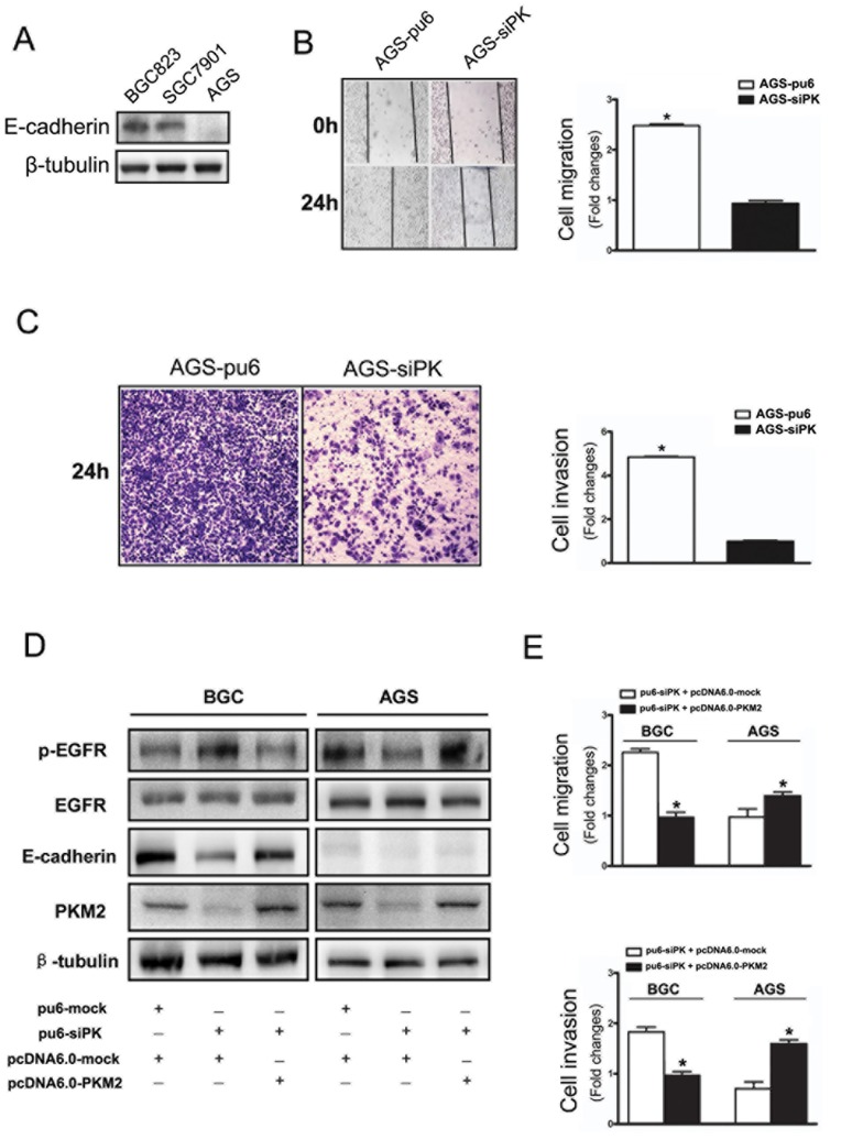
Figure 3. Depletion of PKM2 attenuated the motility of AGS cells and the functional changes after rescuing PKM2 in gastric cancer cell lines.
(A) E-cadherin expression levels were detected by immunoblot analysis in BGC823, SGC7901 and AGS cells. (B) A cross-shaped wound was created in the monolayer, and the AGS stable cells were cultured for an additional 24 h with EGF (100 ng/ml). Representative images of the wounded region are shown. The results of the migration assay are also shown as graphs (*p<0.05). (C) The invasion potential of AGS stable cells was assessed using the BD transwell chamber assay with 100 ng/ml EGF in the lower chamber for 24 hours. The cells that migrated to the lower side of the filter were stained, photographed, and counted. (D) The expression of p-EGFR, E-cadherin in the PKM2 rescuing experiments with stably transfected method by using over-expression plasmid vector pcDNA6.0-mock and pcDNA6.0-PKM2 in BGC823 and AGS cells which stable knockdown PKM2. (E) The functional changes of cell migration and invasion after PKM2 rescuing. The data are expressed as the mean ± SD from three independent experiments (*p<0.05).

