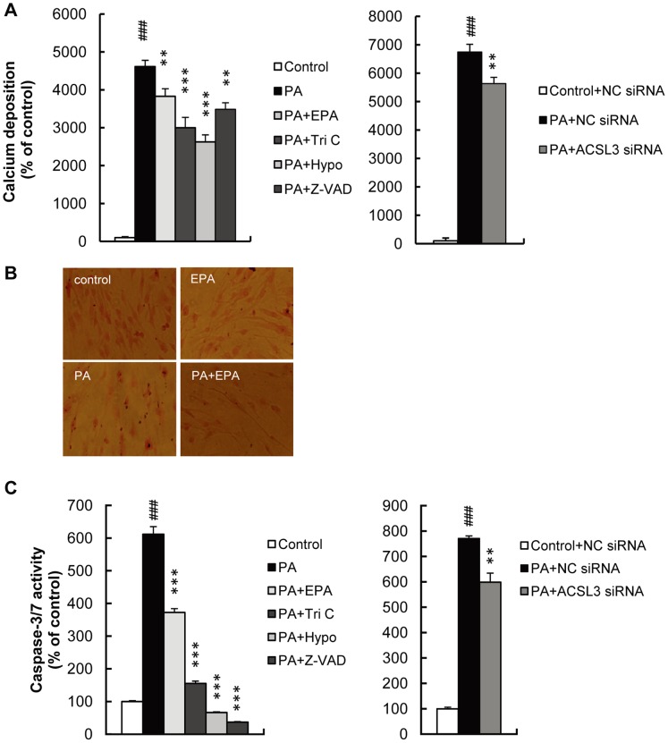
Figure 6. High-concentration PA induced calcium deposition and caspase activation in HASMC.
HASMC were treated with PA (1 mM) for 1 day with or without EPA (15 µM), Tri C (2 µM), Hypo (100 µM) or Z-VAD-FMK (Z-VAD; 100 µM). ACSL3 siRNA were transfected before PA treatment. A, Calcium deposition was determined by the methylxylenol blue method and normalized to protein content. B, Calcium deposition was detected using von Kossa-staining. C, Caspase-3/7 activity was determined as described in Materials and Methods, and normalized to protein content. All values are presented as the mean ± S.E. (n = 3–14). ###p<0.001 vs. control or control+NC siRNA, **p<0.01, ***p<0.001 vs. PA or PA+NC siRNA.

