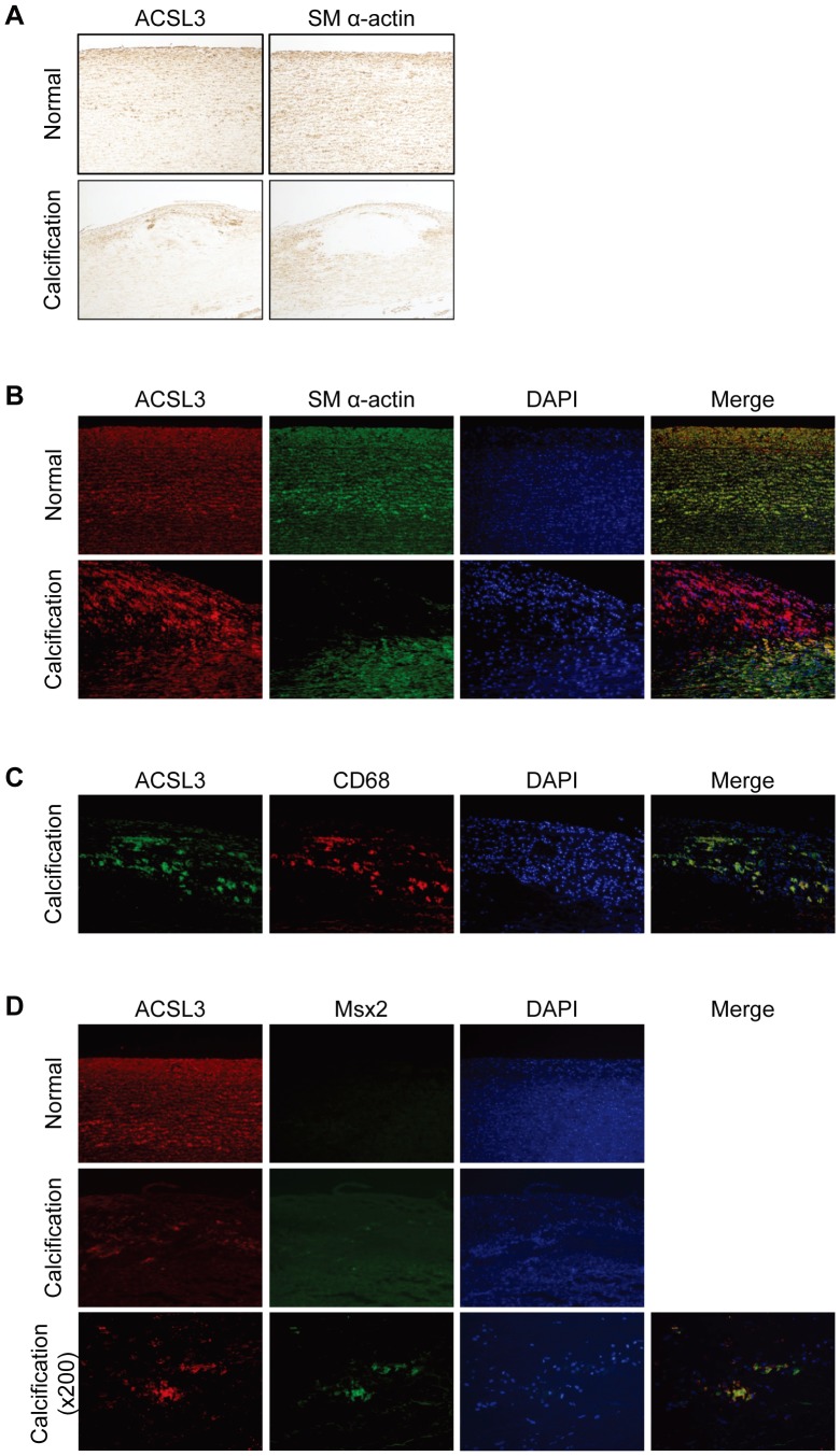
Figure 7. The expression of ACSL3 in normal and calcified human carotid artery.
A, Immunohistochemistry for ACSL3 and SM α-actin in normal (upper panel) and calcified human carotid artery (lower panel). Magnification ×100. B to D, Immunofluorescent labeling for ACSL3 (red or green), (B) SM α-actin (green), (C) CD68 (red) or (D) Msx2 (green) and DAPI (blue) in normal and calcified human carotid artery. Magnification ×100 and ×200 (only calcified human carotid artery in D).

