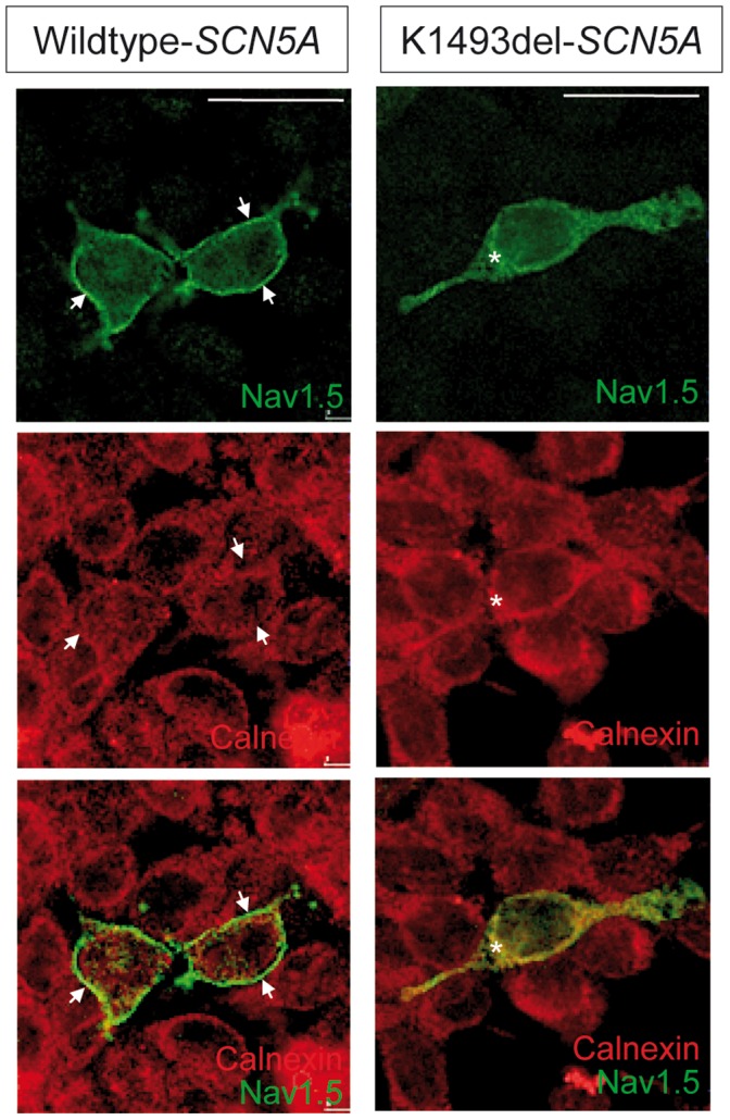
Figure 5. Sodium channel membrane expression in wild-type and mutant 1493delK SCN5A-transfected HEK293 cells.

Confocal immunofluorescence of the a-subunit of cardiac sodium channel (NaV1.5) and the endoplasmic reticulum transmembrane protein calnexin in HEK293 expressing WT (left) and mutant 1493delK (right) sodium channels. Top and middle panels show staining with anti-NaV1.5 (green) and anti-calnexin (red) respectively. Bottom panels show overlay of red and green channels of double staining with anti-NaV1.5 (green) and anti-calnexin (red) antibodies. Membrane labeling for NaV1.5 is observed as a clearly distinguishable green rim surrounding the intracellularly located calnexin (red) in WT SCN5A transfected HEK293 cells, whereas mutant 1493delK SCN5A transfected HEK293 cells do not show clear cell-surface labeling, but mostly cytoplasmic NaV1.5 staining. Scale bars indicate 25 µm.
