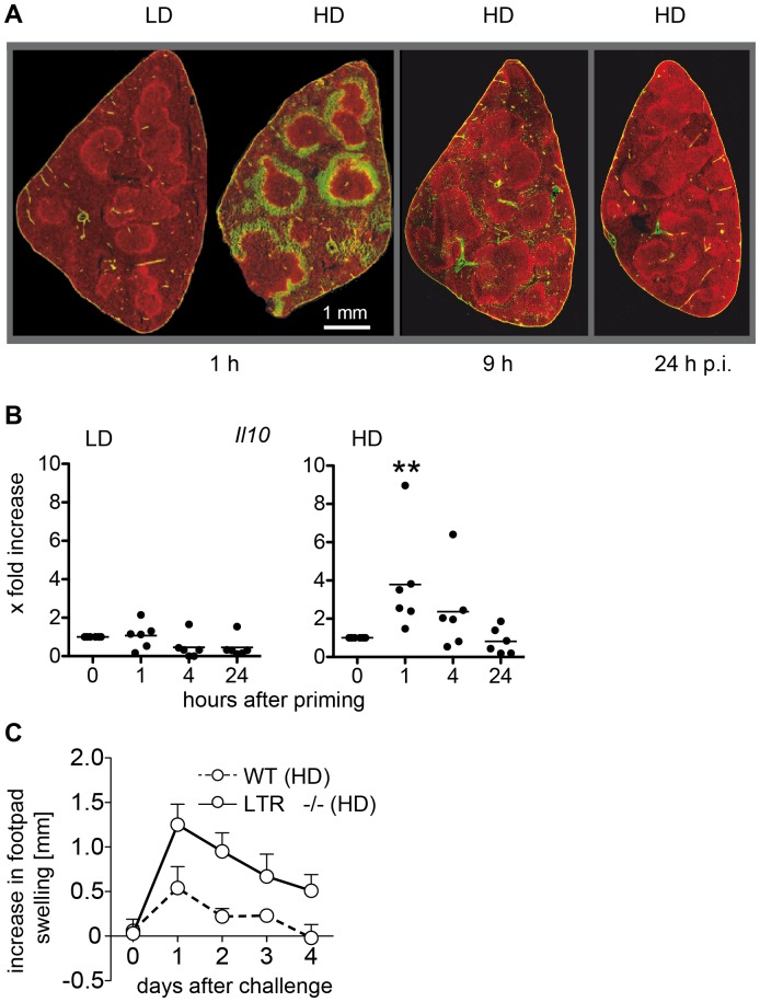
Figure 5. An intact MZ is required for the induction of Th2 cells.
Mice were primed with a LD or HD of SRBC. SRBC were labeled with CFSE before injection (green) and spleens were removed 1 h, 9 h and 24 h after LD and HD priming. Histological sections were prepared and the MZ were visualized by staining with Alexa 555-labelled antibodies against Moma-1, which is specific for metallophilic macrophages (red) [37] (n = 3, one typical section is shown) (A). The MZ was isolated by laser-microdissection at indicated time points and mRNA expression of Il10 as marker for activated MZ B cells was analyzed. Each circle represents one animal (n = 6) (B). Significant differences in the expression of Il10 between primed mice compared to the controls are shown (**p<0.01, Mann-Whitney-U-test, n = 6). Mice were primed with a HD of SRBC. One footpad was challenged with SRBC 5 days after priming. The footpad thickness was measured between 1 and 4 days after challenge of wild type and LTβR−/− mice (C). Data are means ± SD. *indicate significant differences in footpad thickness compared to control mice (**p<0.01, Mann-Whitney-U-test, n = 6). Abbreviations: HD, high dose (109); LD, low dose (105).

