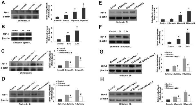
Figure 6. Western blotting analysis of the expression of RIP-1.
The expressional level of RIP-1 increased significantly in C6 (A) and U87 (E) glioma cells when shikonin concentration was elevated. The expression of RIP-1 was up-regulated markedly when the incubation of C6 (B) or U87 (F) glioma cells with shikonin was elongated from 1.5 h to 3.0h. Pretreatment with Nec-1 attenuated the increased expression of RIP-1 caused by shikonin either at lower or higher concentration in C6 (C) and U87 (G) glioma cells. Pretreatment with NAC made the increased level of RIP-1 decrease in both C6 (D) and U87 (H) glioma cells when compared with shikonin group. *: p<0.01 versus control group; #: p<0.01 versus shikonin treated group.

