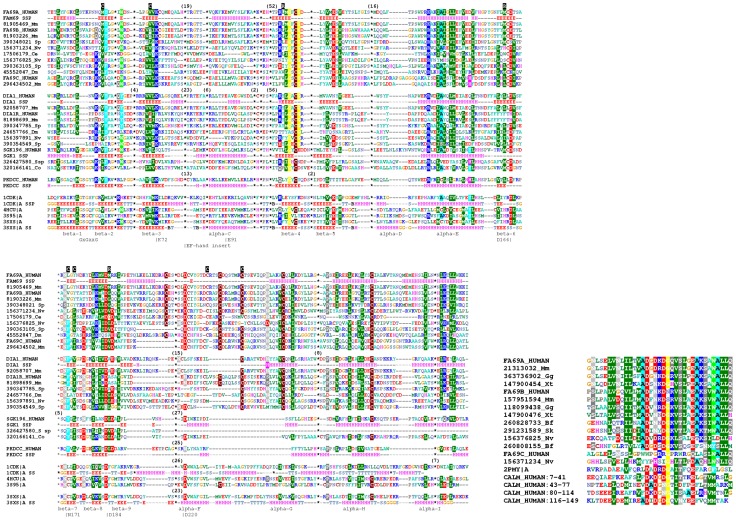
Figure 2. Multiple sequence alignments of selected FAM69 proteins.
Left: alignment of the kinase domain, covering the region 83–422 of FAM69A. The sequences are aligned using Promals3D (see Methods). Secondary structure prediction for human shown for selected proteins, for the solved structures actual secondary structure shown. Secondary structure elements named as in PKA [87]. Locations of predicted key catalytic residues shown, in standard PKA numbering (e.g. D166), as well as the ATP-binding loop (GxGxxG). SwissProt identifiers shown for human sequences, otherwise, NCBI GI identifiers shown together with abbreviations of species names: Sp: sea urchin Strongylocentrotus purpuratus, Nv: sea cucomber Nematostella vectens, Dm: fruit fly Drosophila melanogaster; Mm: Mus musculus; Ce: Caenorhabditis elegans, Co: Capsaspora owczarzaki; S sp: Salpingoeca sp. ATCC 50818. Also shown selected close kinase homologues (PKDCC and SGK196) as well as sequences of selected kinases of known structures. Numbers in brackets indicate numbers of residues omitted from the alignment (shown only for the 1 cdk, 3 sxs structures, and for human FAM69A, DIA1, PKDCC and SGK196 sequences). R and C characters on black background above the alignment indicate the regulatory and catalytic spine residues, respectively [22]. The location of the the EF-hand motif shown, the motif itself is excised from the alignment and shown on the right. Right: alignment of the EF-hand region (corresponding to the region 165–199 of human FAM69A). Also shown EF-hand regions of human calmodulin and the 2PMY region used for model building.

