Fig. 7.

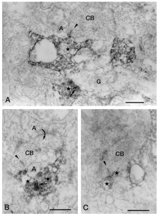
Vertical sections through the IPL stained for Y1 immunoreactivity. A: Y1-immunoreactive amacrine cell process (star) forms a postsynaptic dyad with an unlabeled amacrine cell process (A) at the ribbon synapse (arrowhead) of a CB in stratum 4 of the IPL. In the lower part of the figure, a labeled amacrine cell processes (star) that is making synaptic output (arrow) onto a ganglion cell dendrite (G) can be seen. B: A CB, postsynaptic (curved arrow) to an unlabeled amacrine cell process (upper A), makes a ribbon synapse (arrowhead) onto a postsynaptic dyad consisting of a labeled amacrine cell process (left star) and an unlabeled amacrine cell process (lower A) in stratum 4 of the IPL. The labeled process receives synaptic input (arrow) from another labeled amacrine cell process (right star). C: Two labeled amacrine cell processes (stars) form a postsynaptic dyad at the ribbon synapse of an axon terminal of a CB) in stratum 4 of the IPL. IPL, inner plexiform layer; CB, cone bipolar axon terminal. Scale bar = 0.5 μm.
