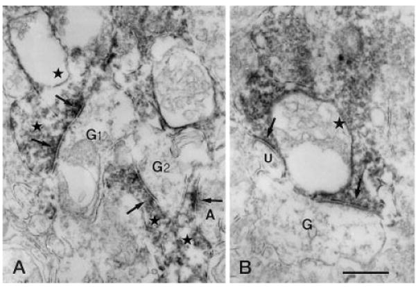
Fig. 8.

Vertical sections through the IPL of a rat retina processed for Y1 immunoreactivity. A: G1 is postsynaptic (left, upper arrows) to two immunoreactive amacrine cell processes (left, upper stars) in stratum 2 of the IPL. Another ganglion cell dendrite (G2) receiving synaptic input from an immunoreactive amacrine cell process (center star) is seen. In the right lower corner of the figure, an immunolabeled amacrine cell process (right, lower star) receives synaptic input (right, lower arrows) from an unlabeled amacrine cell process (A). B: Immunoreactive amacrine cell process (star) makes output synapses (arrow) onto an unlabeled G and an unidentified process (U) in stratum 4 of the IPL. G,G1,G2, ganglion cell dendrite; IPL, inner plexiform layer. Scale bar = 0.5 μm in B (applies to A,B).
