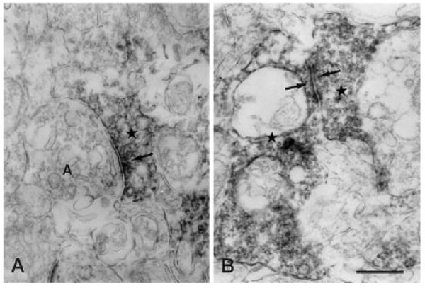
Fig. 9.

Vertical sections through the IPL of a rat retina processed for Y1 immunoreactivity. A: Immunoreactive amacrine cell process (star) makes an output synapse (arrow) onto an amacrine cell process (A) in stratum 2 of the IPL. B: Synaptic contact (arrows) between two immunoreactive amacrine cell processes (stars) is seen in stratum 4 of the IPL. IPL, inner plexiform layer. Scale bar = 0.5 μm in B (applies to A,B).
