
Figure 2.
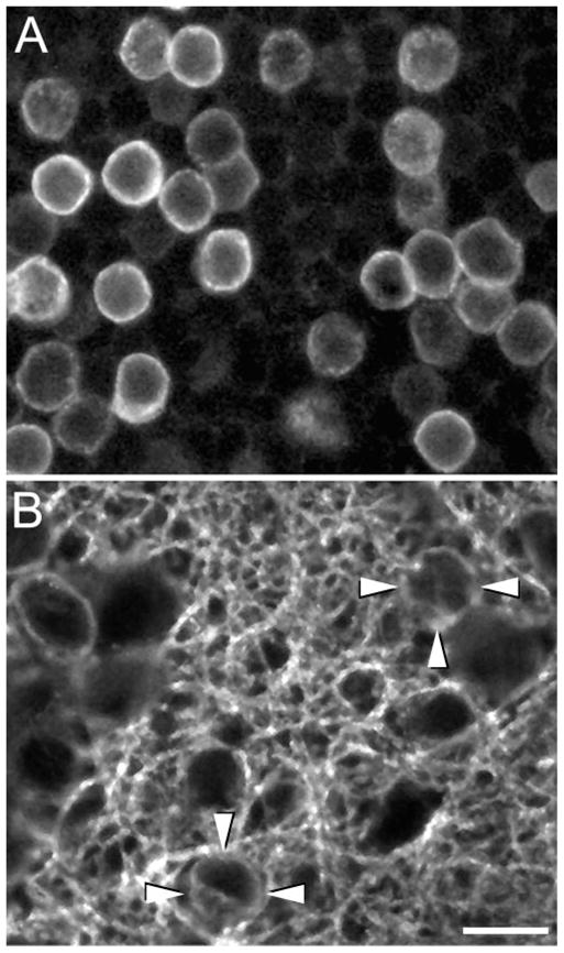
Confocal images (1 μm-thick digital sections) of horizontal sections of a monkey retina showing GAT-1 immunoreactive somata in (A) the proximal INL and (B) the IPL. GAT-1 immunoreactivity outlines the cell somata, indicating that GAT-1 is localized at or near the plasma membrane. (B, arrowheads) Two GAT-1-immunoreactive interstitial cell bodies in the IPL. Scale bar, 10 μm.