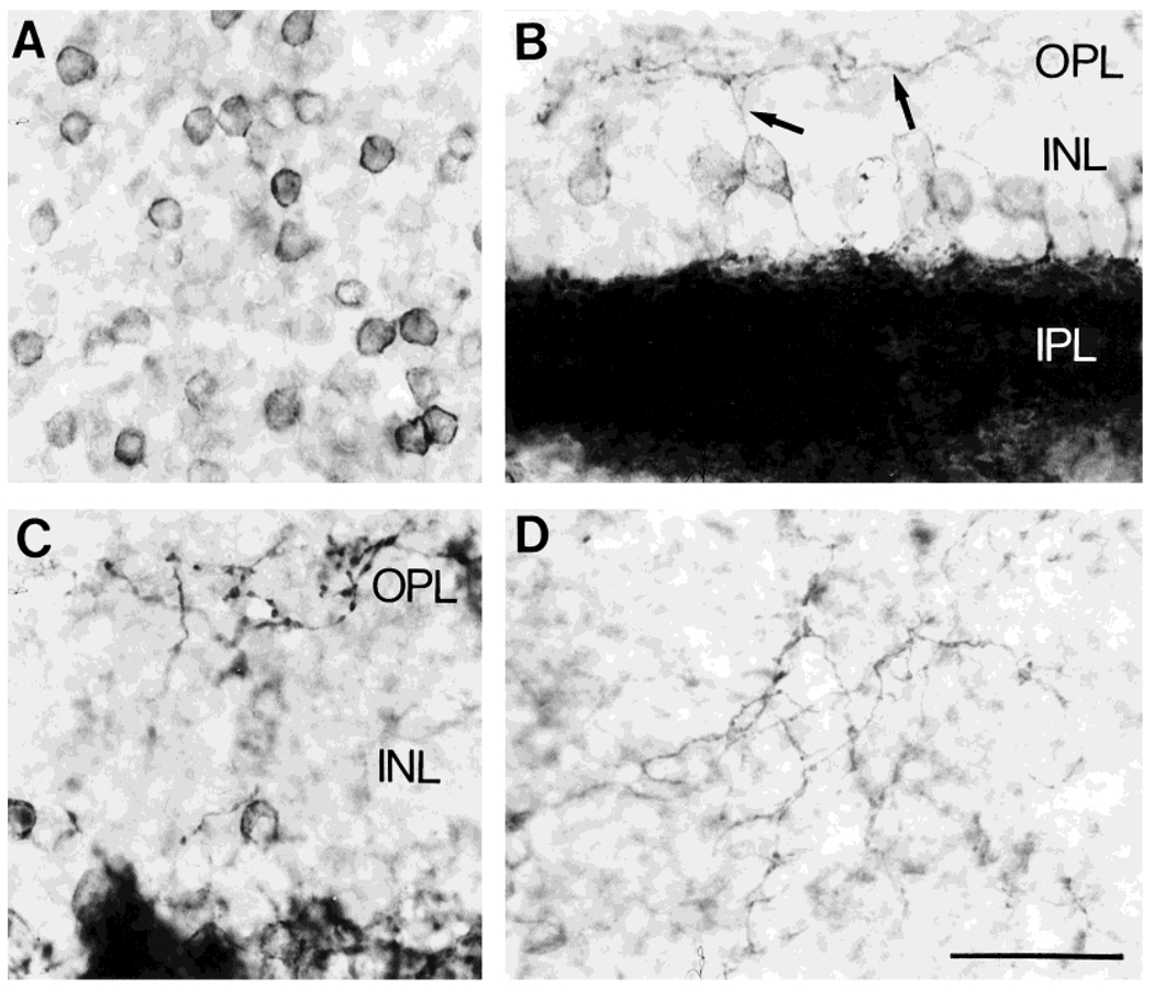
Fig. 2.
NK1 immunoreactivity in retinal sections cut parallel to the vitreal surface. Avidin-biotin-peroxidase technique. A: NK1-IR cell bodies in the INL. B,C: Photomicrographs taken from regions where the section was cut in an oblique plane, so that different retinal layers are present in the same section. Note NK1-IR processes (arrows) innervating and arborizing in the OPL. D: Section through the OPL showing the NK1-IR plexus. Scale bar = 30 µm.

