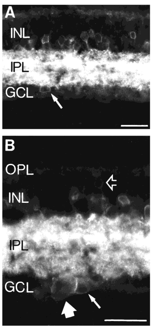
Fig. 3.
A: An NK1-IR cell (arrow) located in the GCL. The soma size of this cell is similar to that of NK1-IR cells in the INL, and it is likely to be a displaced amacrine cell. B: Higher magnification photomicrograph showing a putative NK1-IR displaced amacrine cell (small arrow) together with a larger NK1-IR cell in the GCL (large arrow), which is likely to be a ganglion cell. The open arrow points to a very faint NK1-IR cell in the distal INL. The location and morphology of this latter cell resemble those of a bipolar cell. Scale bars = 30 µm.

