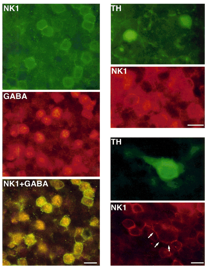
Fig. 4.
Retinal sections cut parallel to the vitreal surface and processed by double-label immunohistochemistry by using antibodies directed against NK1 and γ-aminobutyric acid (GABA), and NK1 and tyrosine hydroxylase (TH). Left: NK1 immunoreactivity is shown in green; GABA immunoreactivity is shown in red; NK1+GABA shows the simultaneous visualization of NK1 (green) and GABA (red-orange) immunoreactivities in the same preparation with a dual-band filter. Note that most GABA-IR cell bodies also display NK1 immunoreactivity, which is confined to their plasma membrane. In addition, some NK1-IR cell bodies do not contain GABA immunoreactivity, and some GABA-IR cells do not express NK1 immunoreactivity. Right: TH (green) and NK1 (red) immunoreactivities visualized with fluorescein and rhodamine filters, respectively. Top two figures show two small TH-IR cell bodies displaying NK1 immunoreactivity at their cell surface; bottom two figures illustrate the colocalization of TH and NK1 immunoreactivities in a large cell (arrows point to the NK1 cell surface staining of such cell). Scale bars = 10 µm.

