
Figure 5. Tau is expressed in neural stem cells, neural progenitor cells and migrating neuroblasts.
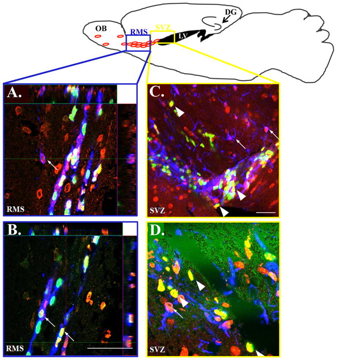
TOP: Schematic presentation showing the neurogenic niches in a sagittal slice through the mouse brain: the region outlined by the blue box is representative of the area from which confocal RMS (A,B) images were taken and the yellow box represents the region corresponding to the SVZ images (C,D). Confocal imaging of immunolabeled brain sections of APPswe/PS1ΔE9 mice shows that tau co-localizes with doublecortin (small arrows; A,B), BrdU (A–D big arrows in C,D) and GFAP (small arrows; C,D). Tau, red (A–D); BrdU, green (A–D); doublecortin, blue (A,B); GFAP, blue (C,D). Scale bar= 50μm