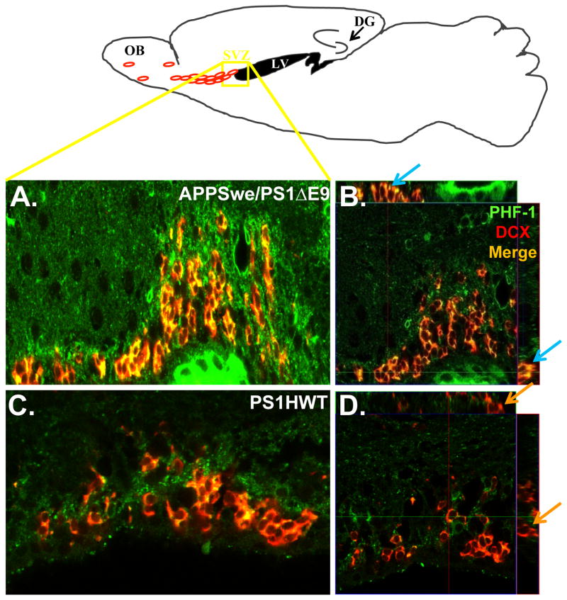
Figure 6. PHF-1 is expressed in neuroblasts in the subventricular zone of APPswe/PS1ΔE9 mice.
Upper Scheme: Schematic presentation of the area in the SVZ from which confocal images were taken. Left panel: PHF-1 expression is pronounced in DCX+ neuroblasts in the SVZ of APPswe/PS1ΔE9 (A) but hardly in PS1HWT (C) mice at 6 months of age. Right panel: High power orthogonal image showing strong co-localization of PHF-1 (green) and DCX (red) in the SVZ of APPswe/PS1ΔE9 (B) and no co-localization in PS1HWT mice (D). Blue arrows represent clear co-localization in the orthogonal view of APPSwe/PS1ΔE9 sections while the orange represent DCX+ cells in the PS1HWT SVZ that are clearly not PHF-1+. Scale bars= 50μm.

