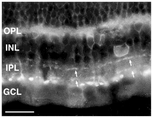
Fig. 5.
The sst2A receptor-immunoreactive amacrine cells were sparsely occurring. Immunoreactive cell bodies were located at the border of the inner nuclear layer (INL) and the inner plexiform layer (IPL). They were likely to be multistratified and gave rise to the processes in laminae 2 and 4 of the IPL. In transverse sections, these processes in laminae 2 and 4 were not continuous. OPL, outer plexiform layer; GCL, ganglion cell layer. Scale bar = 25 μm.

