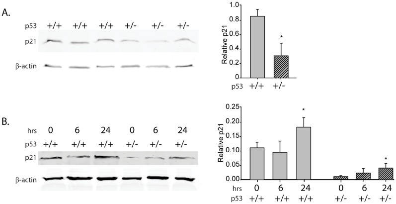
Figure 2. p21 protein in Wnt-1 p53+/+ and Wnt-1 p53+/− tumor suspensions.
A, p21 protein by immunoblot in Wnt-1 p53+/+ and Wnt-1 p53+/− tumor cell suspensions (approximately 1 million cells) and B, in response to UVC DNA damage after 0, 6 and 24 hours. Significant differences in protein levels are indicated by an asterisk on the densitometry plots; P≤0.05.

