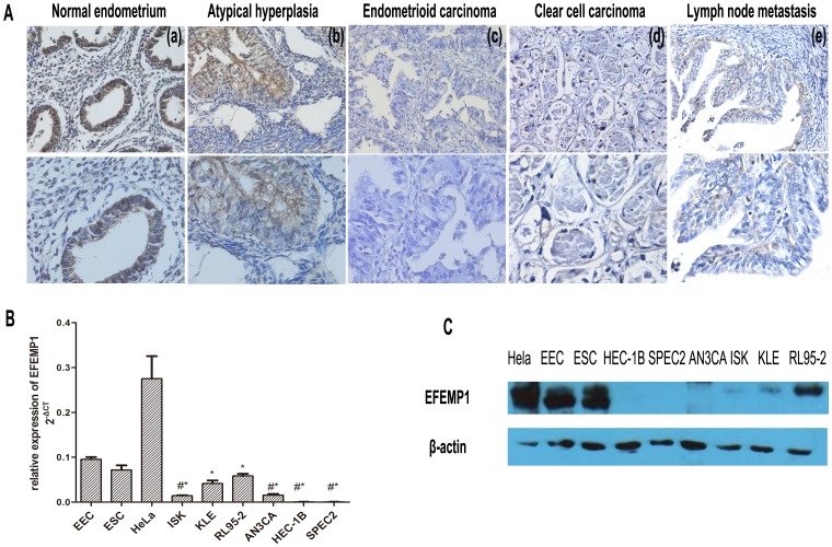
Figure 1. Differential EFEMP1 expression in normal endometrium and endometrial carcinoma.
A: Immunochemistry of EFEMP1 in endometrial tissues. (a) normal endometrium with strong staining, (b) atypical hyperplasia with moderate staining, (c) endometrioid carcinoma with no staining, (d) clear cell carcinoma with no staining, and (e) serous endometrial carcinoma with lymph node metastasis. Magnification:×200 (above) and×400 (below). B: Down-regulation of EFEMP1 mRNA expression in EC cell-lines as compared with EEC (endometrial epithelial cell). ESC is the abbreviation of endometrial interstitial cell. HeLa cells were used as a positive control. The experiment was repeated twice, and each experiment was done in triplicate. * P<0.05, #*P<0.01. Arithmetic means of the data (bars) and SD (error bars) are shown. C: EFEMP1 expression in various endometrial cancer cell-lines as determined by Western immunoblotting analysis.

