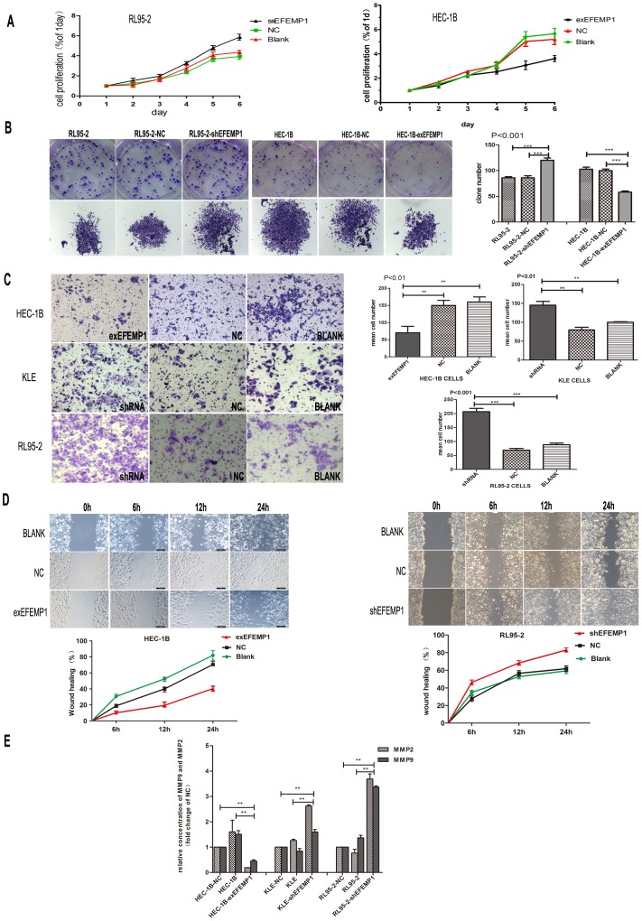
Figure 5. Effect of EFEMP1 on endometrial carcinoma cell proliferation, migration and invasion.
A: Knockdown of EFEMP1 promoted cell growth in RL95-2. However cell proliferation was lower in HEC-1B-exEFEMP1 cells compared with negative control and HEC-1B. B: Plate colony formation assay was used to measure cell proliferation of EFEMP1 in HEC-1B-exEFEMP1 and RL95-2-shEFEMP1 and negative control. C: Representative images and statistical plots of Transwell assays in HEC-1B cells, which were stably transfected with exEFEMP1and vector control; RL95-2 cells and KLE cells transfected with shEFEMP1 and vector control (x200 magnification). The data were described as arithmetic mean ± SD of three independent experiments. D: Wound healing assays of cells expressing the vector control and exEFEMP1 in HEC-1B; and RL95-2 cells showing dampened expression of EFEMP1and vector control. Typical images were captured at the precise time of wounding at 0, 6, 12 and 24 h (original magnification×100). The percent of wound closure was measured in at least three randomly selected regions (mean ± SD). E: MMP-2 and MMP-9 secretion was decreased significantly in HEC-1B transfected with exEFEMP1 and increased in KLE and RL95-2 transfected with shEFEMP1cells. Data are expressed as mean concentrations ± SD of three independent experiments. * P<0.05, **P<0.01, ***P<0.001.

