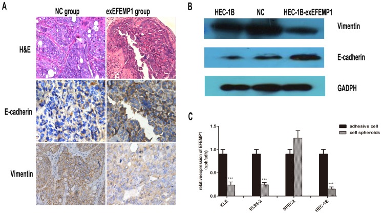
Figure 6. The effect of EFEMP1 on E-cadherin and vimentin expression.
A: The nude mouse tumor tissues were paraffin embedded and tumor slides were stained with hematoxylin & eosin (×400), antibody of E-cadherin(× 400) and vimentin (×400). B: Western immunoblot analysis of the changes in expression of E-cadherin and vimentin in HEC-1B cells transfected with exEFEMP1 and vector. C: Cell spheroids cultured to mimic metastatic cancer cells. qRT-PCR analysis of the changes in EFEMP1 expression in various EC cells. Data are expressed as the mean; bars, ±SD, and normalized to adherent cells from three independent experiments. ***P<0.001.

