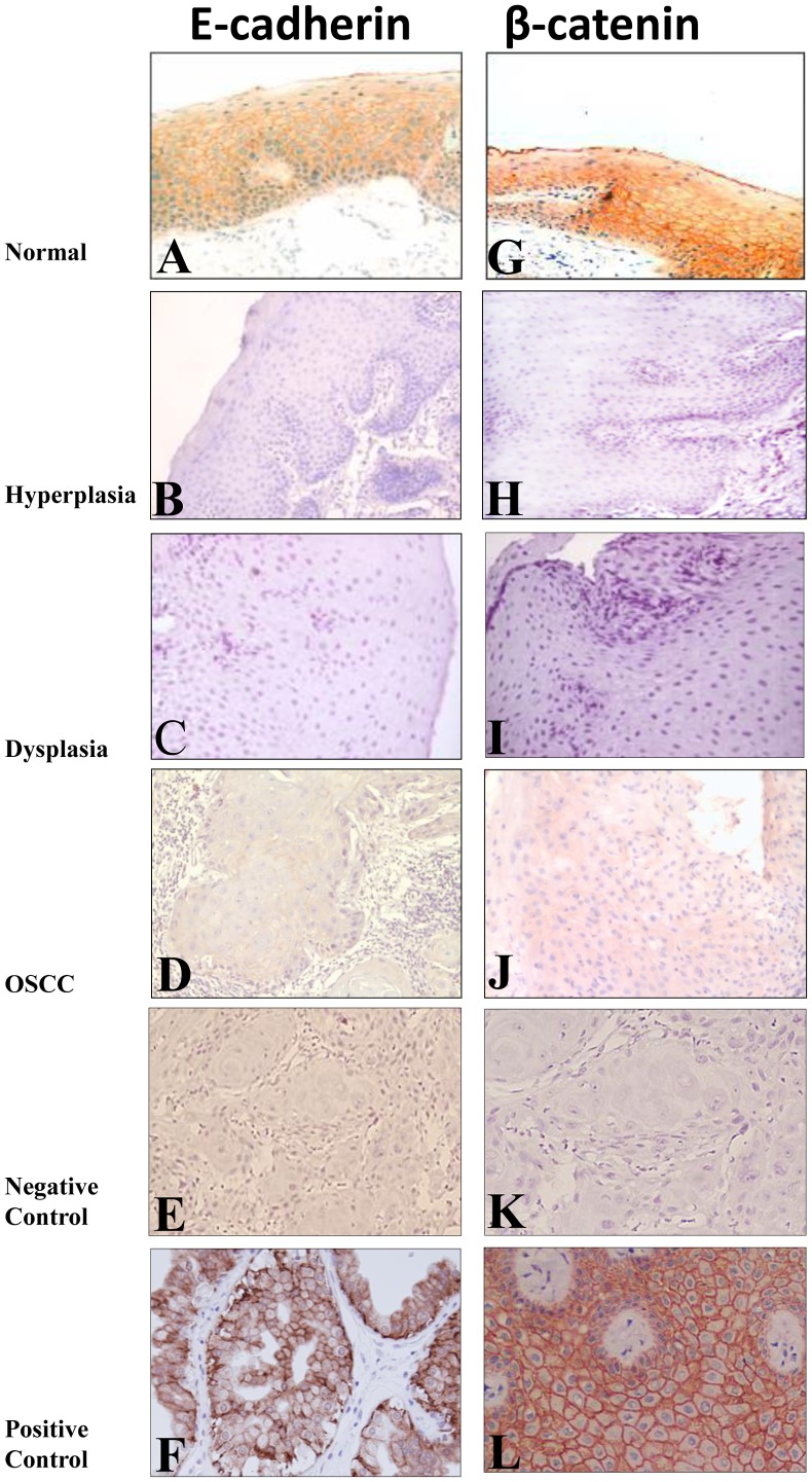
Figure 1. Representative oral tissue sections immunostained for E-cadherin and β-catenin.
Histologically normal tissue sections showing membranous immunoreactivity for E-cadherin (A), and β-catenin (G) proteins. Hyperplastic (B), dysplastic (C), OSCC (D) tissue sections depicting loss of membranous staining for E-cadherin. Hyperplastic (H), dysplastic (I), OSCC (J) tissue sections depicting loss of membranous staining for β-catenin. In negative controls, for E-cadherin (E) and β-catenin (F), the primary antibody was replaced by non-immune IgG of the same isotype to ensure specificity. Breast cancer tissue sections used as positive control showed membrane staining for E-cadherin (K) and β-catenin (L) proteins. Original magnification X 200.

