
Figure 1.
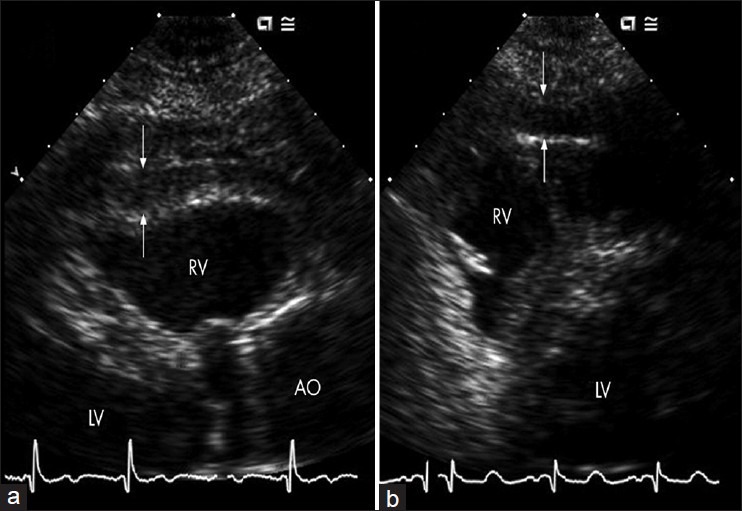
Transthoracic echocardiogram showing a large area of epicardial adipose tissue (white arrows) on free wall of right ventricle (a and b). RV: right ventricle, LV: Left ventricle, Ao: Aorta
Official websites use .gov
A
.gov website belongs to an official
government organization in the United States.
Secure .gov websites use HTTPS
A lock (
) or https:// means you've safely
connected to the .gov website. Share sensitive
information only on official, secure websites.
Transthoracic echocardiogram showing a large area of epicardial adipose tissue (white arrows) on free wall of right ventricle (a and b). RV: right ventricle, LV: Left ventricle, Ao: Aorta