
Figure 5.
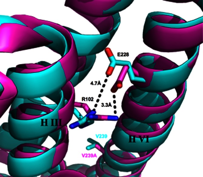
Superposition of helices III and VI of the inactive thermostabilized A2AR mutant (containing the V239A mutation) structure (pink, Pdb accession code 3PWH) and the inactive A2AR bound to an antibody (blue Pdb, accession code 3VG9). The V239 or V239A and E228 residues located on helix VI and the R102 residue located on helix III are shown as stick models. The distance between the charged interacting groups of E228 and R102 of the ionic lock in each case are indicated by the dotted lines.