
Figure 9.
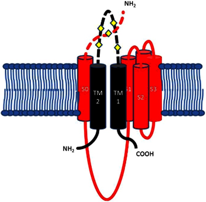
The potential extracellular binding site between the extracellular loop of the β1 subunit and the Nterminal of S0 domain of the α subunit. β1 TM1 and TM2 are black, with TM2 next to S0. Residues 16–20 of the extracellular N-terminal segment preceding S0 have been modelled as a random coil, crossing over the S3–S4 loop. This diagram represents the potential interface and binding site between one of the β1 subunits and one of the four α subunits making up a BKCa channel. The diamonds represent the extracellular cysteines.