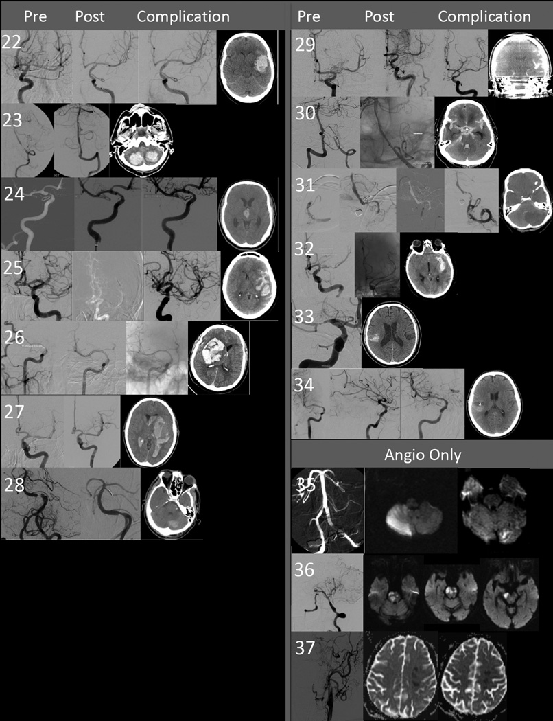
Figure 2.
Composite figure of pre and post-angioplasty and stenting angiogram, and brain imaging demonstrating the location and extent of hemorrhage or ischemic injury for each individual patient. The number corresponds to the patient number in tables 1, 3, and 4. Descriptions of the procedure and relevant findings on the images is found in the table under the description of the headings “timing and details of procedural” and “Post-procedural details”.

