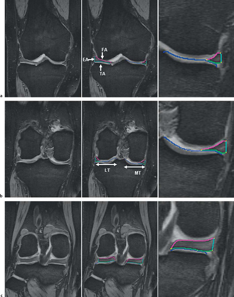
Fig. 1.
Coronal MRI of the right knee without segmentation (left), with segmentation of the MM and LM (middle), and with the segmentation of the MM zoomed (right). The tibial plateau area (ACdAB) is shown in blue, the TA in green, the FA in magenta, and the EA in turquoise. a MRI at the level of the anterior horns. b MRI showing the meniscal body. c MRI at the level of the posterior horns.

