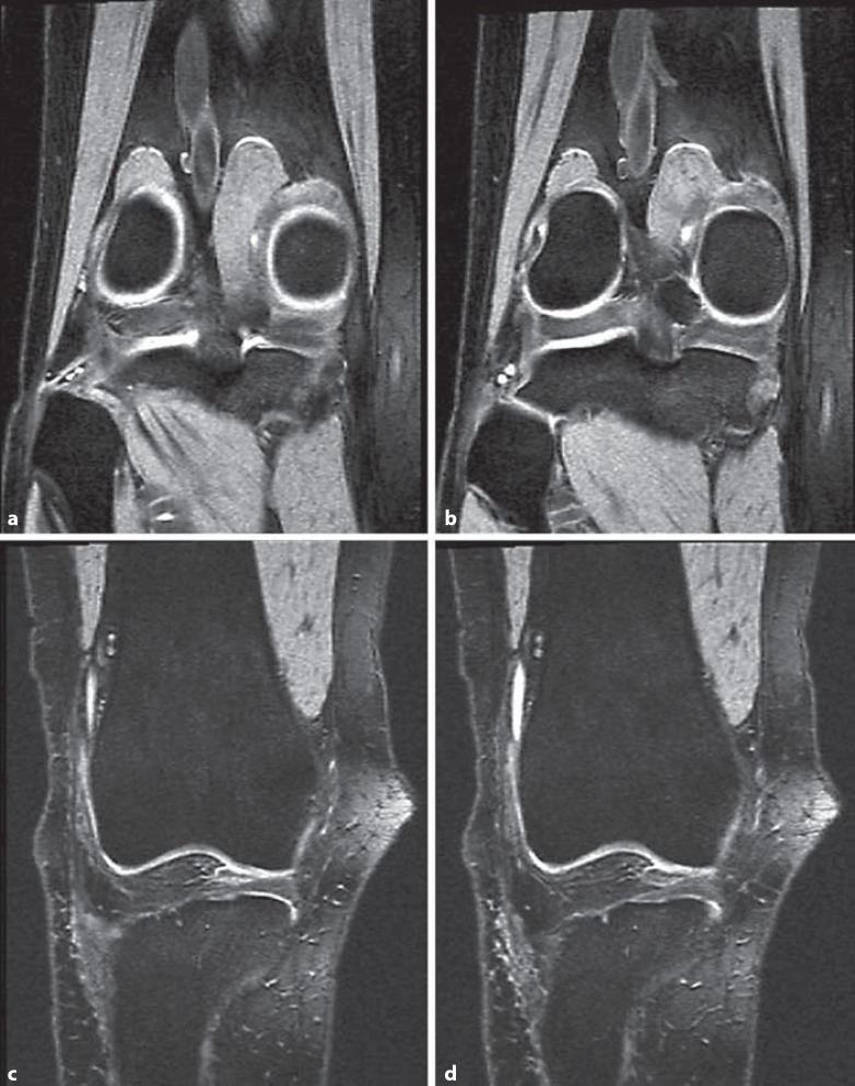
Fig. 2.
Coronal MRI showing the anterior and posterior limits of meniscus segmentation. a MRI slice posterior to the MM, which was not segmented, because the MT plateau or the MM could not be reliably delineated. b First MRI slice in which the MM and MT plateau was segmented. Segmentation is not shown for better clarity. c Last MRI slice in which the MM and MT plateau was segmented. Segmentation is not shown for better clarity. d MRI slice anterior to the MM, which was not segmented, because the MT plateau or the MM could not be reliably delineated.

