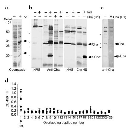
Figure 2.
Reactivity of chagasic sera. (a) Coomassie blue staining shows the expression of recombinant Cha in induced (+) or not induced (–) bacterial lysates of E. coli. Arrows indicate Cha and sCha proteins. (b) Western blot analysis of the same lysates with normal rabbit serum (NRS) and anti-Cha and its competition with the immunizing Cha peptide (R1) is shown. The same lysates were tested with normal human serum (NHS) and with chagasic human serum (Ch+HS). Arrows indicate 34-kDa (Cha) and 20-kDa (sCha) proteins. (c) Western blot analysis of a Jurkat lysate with the anti-Cha Ab. Recognition of specific bands of Cha and sCha is denoted by arrows. (d) Mapping of the sCha epitope recognized by human chagasic sera. The reactivity of six human chronic chagasic sera against 25 overlapping ten-aa peptides spanning sCha was assayed using ELISA. Five sCha-positive sera (filled circles) and one negative sera (open circle) are shown. Peptide 1 corresponds to the R3 sequence.

