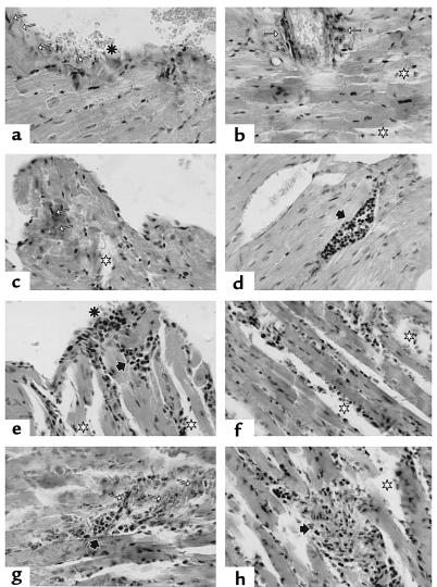
Figure 7.
Histologic study of heart tissue sections from T. cruzi and adoptive transferred mice. (a–d) CBA/J mice in chronic phase of T. cruzi infection. (a) endocarditis (asterisk); note the increase of collagen fibers perpendicular to the tissue surface (white arrows); (b and c) strong presence of perivascular and intermyofibrillar collagen fibers (white arrows); disperse area of edema (white stars); (d) vascular dilatation with lymphocytic thrombus (black arrows). (e–h) Cardiac tissue section of CBA/J mice 2 months after transfer of T cells from chronically infected CBA/J mice, as above. (e) limited myocarditis zone (asterisk) with subendocardiac lymphocytic cumulus (black arrow): (e, f, and h) general edema (white stars); (g) persistence of collagen fibers (white arrow) with strong lymphocytic cumulus in the myocardium (black arrow). van Gieson staining was done in all the sections except for sections e and h, where a hematoxylin and eosin staining was performed. ×400.

