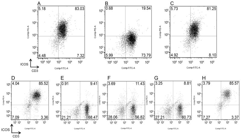
FIGURE 2. ICOS specificity and expression on T-cells mediated by anti-CD3/CD28 or alloantigen stimulation.
A) Gated CD3+ PBMC stained with anti-caICOS-PE. B) Gated CD3+ PBMC stained with a mixture of anti-caICOS-PE (5 μg/ml) and ICOSmurineIg fusion protein (500 μg/ml). C) Gated CD3+ PBMC stained with a mixture of anti-caICOS-PE (5 μg/ml) and the negative control fusion protein CTLA4-Ig (500 μg/ml). Flow cytometry analysis of ICOS expression on CD3+ cells gated from PBMC after culture for 7 days on tissue culture dishes coated with anti-CD3 mAb coated at 10 ug/ml (D), medium only (E), anti-CD3 mAb at 1 μg/ml (F), anti-CD28 mAb at 10 μg/ml (G), or the combination of anti-CD3 (1 μg/ml) and anti-CD28 (10 μg/ml) (H).

