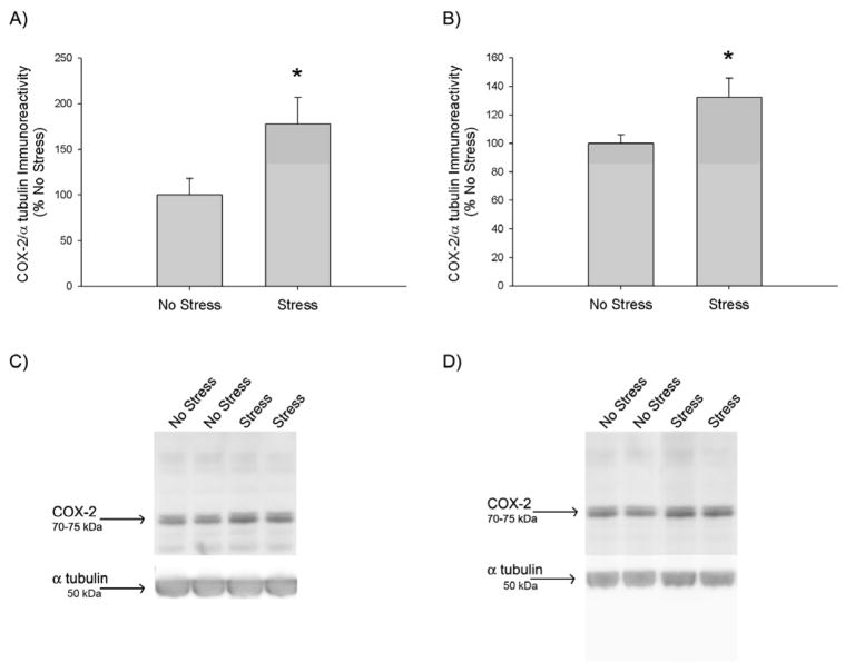
Figure 1.
Effects of CUS on COX-2 protein expression. Rats were exposed to 10 days of CUS or daily handling. On the day after the last stressor, A) hippocampal COX-2 and B) striatal COX-2 immunoreactivity were quantified via Western Blot. CUS significantly increased COX-2 protein expression in the A) hippocampus (*, p<0.05) and B) striatum (*, p<0.05), compared to No Stress, as indicated by a t-test (n=5–6 for each group). Representative Western Blot images of C) hippocampal and D) striatal COX-2 (~72 kDa) and the α tubulin (50 kDa) loading control.

