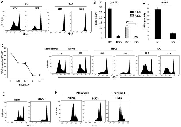
Figure 2. Human HSCs markedly inhibit T cell response.
(A) Human HSCs stimulate low T cell proliferative response. CFSE-labeled PBMC-derived T cells were cultured with irradiated stimulators (allogeneic DC or HSCs) at a stimulator: responder ratio of 1:10 for 5 days. Proliferative response was analyzed by CFSE dilution assay gated in CD4 and CD8 cell populations. (B) Less CD4 and CD8 T cells are generated following HSCs stimulation. Cells in each well were counted at the end of cultures. The numbers of CD4 and CD8 T cells were calculated based on flow analysis. (C) Less IFN-γ is produced by T cells stimulated by HSCs. Supernatant was collected at the end of culture, and the IFN-γ levels were measured by cytometric bead array (CBA, BD biosciences) assay. (D) HSCs inhibit T cell responses. Graded number of irradiated HSCs or DC (as regulators) were added at beginning into a one-way MLR culture in which T cell proliferative response was elicited by allogeneic DC at DC:T ratio of 1:10 for 5 days. T cell proliferative response was examined by thymidine up-take assay (left panel) or by CFSE dilution assay (right panels). (E) HSCs inhibited T cell response elicited by TCR ligation. Irradiated HSCs were added at the beginning into a culture (3 days) of CFSE-labeled T cells (at a ratio of 1:10), proliferation of which were elicited by anti-CD3/CD28 beads. T cell proliferative response was determined by CFSE dilution assay gated in CD3+ cells. (F) HSCs induced T cell suppression is mediated by cell-cell contact. To avoid cell-cell contact, transwell was used, in which irradiated HSCs were separated with CFSE-labeled T cells (stimulated by anti-CD3.CD29 beads). T cell proliferative response was determined by CFSE dilution assay gated in CD3+ cells. The data are representative of three separate experiments.

